


TOP > Report & Column > The Forefront of Space Science > 2011 > Sand of Itokawa
![]()

Configuration of sample particles The size of particles picked up by the optical-microscope image ranges from under 10µm to about 300µm. The lowest limit of size is determined by the resolution of the optical microscopes (i.e., several µm) used. It is relatively easy to handle small particles by changing the applied voltage on the manipulator probe. We can attach the particles to or release them from the probe by controlling the electrostatic force. If the particles are large, however, the gravitational effect emerges. The largest particle that we have ever moved is about 300µm. Although we usually use a single electrostatic- controlled probe, there were cases when we used two (see Fig. 5). We perform optical microscope observation when picking up particles from the quartz glass lid. Except for large particles, however, we were unable to investigate in detail the structure of the particles because of the limits of the microscope's resolution. 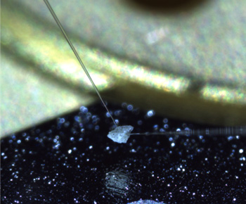
Prior to the detailed analysis, configuration information about each particle was obtained from the SEM-observation in the curation facility. From SEM observation, we confirmed a variety of particle configurations including: ones with a smooth surface; probable chips from a single crystal; multicrystal particles (Fig. 6A, B); ones likely combining many particles with space (Fig. 6C); aggregate of extremely fine particles: and coarse particles wrapped with fine particles. In case of Fig. 6A, the particle looks flat in the SEM back scattered electron image (left), but the optical-microscope image clearly shows that it is an aggregate of many particles. The SEM back scattered electron image in Fig. 6B clearly shows that the particle is an aggregate of minerals with different-composition. There are also many particles with large cracks and complex shapes that were too fragile. In some cases, when we tried to pick them up with the manipulator's electrostatic probe, the particle broke into pieces. Some particles even shattered due to the charge during SEM observation. 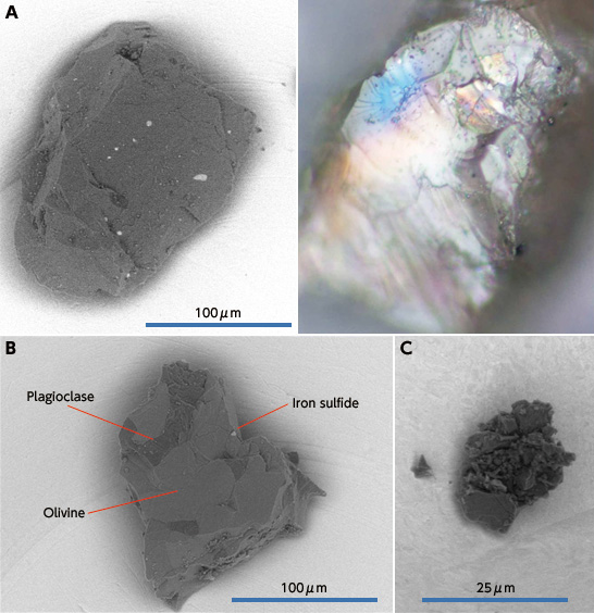
Sedimentary rocks on the earth such as mudstone, sandstone and conglomerate are originally made of mud, sand, and pebbles. They were crushed and compacted to transform into rock. On the other hand, igneous rock was formed in the igneous process, such as volcanoes. Sedimentary and igneous rocks are changed into metamorphic rock due to underground heat and pressure. If sedimentary, igneous and metamorphic rocks are crushed again, they return to mud, sand, and pebbles. I like the expression "Sands of Itokawa." In what stage are the sample particles of Itokawa we see by SEM? Like the earth's sedimentary rocks, will the sands of Itokawa go through the process of compaction and consolidation and transform to meteorites? Or, are the fine particles we obtained chips of rock that compacted in the past like meteorites? Though tiny, the sample particles of Itokawa contain a great amount of information to answer many questions, including the above. Since we have now obtained the materials, it is an exciting challenge for us, humankind, to answer the questions. Akio Fujimura *The tasks Eincluding trial collection of solid-particle test pieces by micro-manipulator, sample recovery by a Teflon spatula, and observation and analysis Ewere conducted by Associate Prof. Tomoki Nakamura (Tohoku University), Prof. Takaaki Noguchi (Ibaraki University), Assistant Prof. Ryuji Okazaki (Kyushu University) and the curation staff.
|
||||||||||